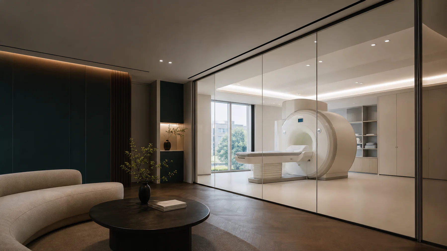
If you have searched for the difference between an MRI and an MRA, the short answer is that they are the same machine doing two different jobs, usually in the same appointment. A brain MRI images the brain itself: the tissue, its structure, anything growing or shrinking where it should not. A brain MRA images the arteries that feed it. One looks at the rooms, the other looks at the plumbing, and on a screening trip you generally want both read together rather than either alone.
That is worth saying plainly, because the search terms people actually type (“mra head scan”, “mri mra brain”, “difference between mri and mra”) suggest a lot of readers arrive unsure whether they are choosing between two tests. Most of the time you are not, and what you are really deciding is whether the scan you book covers the vessels as well as the tissue.
What each one is actually looking at
An MRI of the brain produces a detailed structural map. It is the test that finds a tumour, shows the scarring left by a past small stroke, and picks up the changes that go with a range of neurological conditions. It uses a strong magnetic field and radio waves, not radiation, so the dose is zero. A brain and its vessels together take roughly twenty to thirty minutes on the table.
An MRA reconstructs the arteries from the same physics. Its job is the vessel wall and the vessel lumen: an aneurysm bulging where an artery branches, a narrowing that restricts flow, a malformation that was always there. For anyone with a family history of aneurysm or stroke, the MRA is the part of the appointment that speaks to that history directly, which is why leaving it off a brain scan can quietly remove the reason you booked.
Neither test is a functional or cognitive assessment. A structural scan cannot tell you how well your memory is working, and it does not diagnose early dementia on its own. It shows the anatomy, and a clinician reads that anatomy against the rest of the picture. A scan read without that picture tends to produce more worry than it resolves, which is a theme worth carrying into any imaging decision.
The prices, and where they sit
Because a Korean partner centre has to publish its non-covered (비급여) prices, the standalone figures here can be quoted rather than estimated. A partner centre posts a brain MRI at about ₩319,000 and charges the cerebrovascular MRA again at the same figure, read from its own list on 2026-08-05.
| Scan | Standalone price | What it reads |
|---|---|---|
| Brain MRI | ≈₩319,000 | Brain structure, tumours, evidence of past small strokes. |
| Brain MRA | ≈₩319,000 | The major brain arteries, narrowing and aneurysms. |
| Cheapest programme with both | ≈₩2,000,000 | Hanaro's Gold programme lists brain MRI and MRA together, with a published exam time of four hours. |
Inside a screening programme, the brain scan sits in the top tier, the one that adds an MRI by region. Brain is one of three regions offered, alongside the cervical and lumbar spine. If your reason for scanning is the brain and its vessels, it is worth naming that in the request, because left to the day the MRI region can be assigned to something else, and you can end up leaving without the scan you came for. The full detail of scan time, dose and what it does and does not find sits on the brain MRI and MRA test page.
When the vessel pass earns its place
Not every brain MRI needs its MRA. A structural scan on its own is a reasonable baseline for someone with no particular vascular history who wants to know what their brain looks like today. The MRA earns its place when there is a reason to look at the arteries: a relative who had an aneurysm or a stroke, high blood pressure that has run uncontrolled, or simply the wish to check the one thing a structural scan does not cover. The cost of adding it is one more line on a published list, not a second appointment.
The comparison people reach for next is usually the whole-body MRI, which also passes over the head. That coverage is a general survey, not a dedicated brain study. A whole-body MRI is built to sweep the torso and screen broadly; a brain MRI and MRA use a finer protocol tuned to brain and vessel detail, which is why the two are sold as separate tests rather than one standing in for the other.
The practical version
For most people booking a screening trip, the decision is not MRI or MRA. It is whether the brain scan you are quoted includes the vessels, whether the region is set to the brain rather than left open, and whether the reason you are scanning is written down before you arrive. Get those three right and the difference between the two acronyms stops being a decision and goes back to being what it is: one appointment, two readings, no radiation, and a written report about a week later. If the spine is the other thing on your mind, the spine MRI is priced and scheduled the same way, and the choice between regions is worth making before the day rather than on it.